What is Skin Malignant Melanoma?
Melanoma is a type of skin cancer — one of the most serious types because advanced melanomas have the ability to spread to other parts of the body. Surveillance data predict that more than 62,000 new cases of melanoma will be detected in the US in 2008. The incidence of melanoma is increasing at a rate which is the fastest among all cancers. In areas of high sun exposure the incidence is almost doubling every decade, although is less exposed areas this dramatic raise has been slowing. Causes of melanoma may include increased sun exposure and an increase penetrance of UV rays through a damaged ozone layer. (Melanoma can also develop in the eye, called intraocular melanoma, or rarely in other parts of the body where pigment cells are found. The CIS can provide information about the diagnosis and treatment of intraocular melanoma.) Melanoma begins when melanocytes (pigment cells) gradually become more abnormal and divide without control or order. These cells can invade and destroy the normal cells around them. The abnormal cells form a growth of malignant tissue (a cancerous tumor) on the surface of the skin. Most melanomas will assume a black or dark brown color. Variants of melanomas can nevertheless be not pigmented at all, or assume various colors such as red, purple, or multi-color. Melanoma can begin either in an existing mole or as a new growth on the skin. In fact, only about one tenth to one quarter of melanomas occur in pre-existing skin, with the rest arising in apparently normal skin, predominantly in areas of sun exposure. A doctor or nurse specialist can tell whether an abnormal-looking mole should be closely watched or should be removed and checked for melanoma cells. The purpose of routine skin exams is to identify and follow abnormal moles. Although identified early the cured rate for melanoma approaches 100%, the late stages have a much worse prognosis, with almost 8,000 deaths resulting annually because of this disease.
Who Gets Melanoma?
Skin cancer can be found in people of any age and is one of the more common cancers to find in those younger than 30. The risk of being diagnosed with melanoma increases with age and the average age for it to be found is 61. Those with lighter skin are much more likely to get melanoma than those with darker skin. A major risk factor for most melanomas is exposure to UV rays from sunlight or tanning beds. This is why proper sun protection is so important. Besides damaging rays from the sun, family history could also be a contributing factor to developing melanoma.
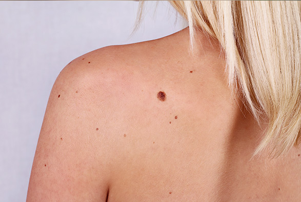
Risk factors for Melanoma
- Family history of melanoma
- Dysplastic nevi
- History of melanoma
- Weakened immune system
- Many ordinary moles (more than 50)
- Ultraviolet (UV) radiation
- Severe, blistering sunburns
- Freckles
- Fair skin
A skin constitution which is sensitive to the sun
Blistering sunburns in early childhood may be the most important risk factor, but the total lifetime exposure may also be important. Melanoma can be produced by ultraviolet exposure in the form of UVB or UVA. Use of tanning beds may result in similar ultraviolet exposure and risks to develop melanoma. Persons who have other types of cutaneous cancers are also at risk of developing melanoma.
Moles and Freckles
(especially if they are multiple, atypycal or “dysplastic”)
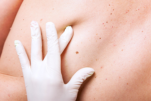
Presence of a large number of pigmented lesions such as freckles or moles carry an increased risk of developing melanoma. The moles are regarded more as a sign of increased risk of melanoma, because the cancer does not always arise in a mole, and it actually may form on apparently healthy skin. The atypical “dysplastic” moles associate a higher risk for melanoma, which can be increased up to 20 times. The “dysplastic mole syndrome” consisting of numerous such nevi is a particularly high risk setting. Features that associated with atypical nevi include a larger size, varitions in in color, and poorly-defined margins. Presence of large congenital nevi associates a risk of malignant transformation into melanoma. Although a complete removal of such lesions can prevent development of melanoma, this can be impractical for those nevi of very large dimensions because of the poor cosmetic results
Type of Skin
Fair skin poses an increased risk because of easy burnig and lack of protection associated with presence of pigment (melanin) into the skin layers.
Personal History of Melanoma
A history of a melanoma harbors a significant risk for developing a second lesion, which may form in up to one quarter of patients, depending also on the presence of other risk factors.
Positive Family History
Approximately one tenth of patients with the malanoma can trace a family member who also had melanoma. The risk is greter if an individual has first-degree relatives affected by melanoma, especially if this occurs under the age of 50, but presence of second-degree relatives is also significant.
Weak Immune System
The immune system can have a diminushed protective ability in response to excessive sun exposure, HIV infection, after chemotherapy or organ transplantation. Certain skin cancers, including the basal cell carcinoma, squamous cell carcinoma, and melanoma are encountered more frequantly in such conditions which produce immunosuppression.
Presence of certain predisposing skin conditions such as Xeroderma pigmentosum, in which there is a defect in DNA repair also enhance the chance of developing melanoma. The National Cancer Institute booklet What You Need To Know About™ Melanoma has more information about risk factors for this disease.
It is important to remember that not everyone who has dysplastic nevi or other risk factors for melanoma gets the disease. In fact, most do not. Also, about half the people who develop melanoma do not have dysplastic nevi, and they may not have any other known risk factor for the disease. At this time, no one can explain why one person gets melanoma while another does not. Research has shown that sun exposure, especially excessive exposure that leads to bad, blistering sunburns, is an important and avoidable risk factor. Scientists are continuing their studies of risk factors for melanoma.
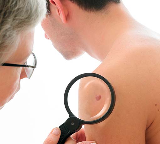
Types of Melanoma
Health professionals distinguish four types of melanoma. The most common is superficial spreading melanoma, which is encountered in almost three quarters of cases. This subtype originates “in situ”, meaning that it develops and extends in the most superficial portion of the skin before starting to grow more deeply (“vertical growth”). Commonly the lesion appears as a discolored patch with suspicious features, such as increasing size, irregular borders, or change in color such as black, round, red, blue, or unstained patches. It is more common in young individuals. Because the superficial pattern of growth may last for up to a few years, this lesion can be diagnosed early, before a “vertical growth phase” develops caring the risk of systemic metastases. Acral lentiginous melanoma is a rare subtype, but is frequently encountered in African-American and Asian patients. It also spreads superficially at the beginning. The initial lesion usually occurs as a dark spot on the palms and soles, or under the nails. Lentigo maligna, also a rare subtype, arise from superficial lesions and commonly keeps a non-invasive pattern of spread for a relatively long period of time before producing deep invasion. It tends to originate in sun-damaged skin, mostly on the head and neck, and frequently appears as brown or black lesion, which is flat or slightly prominent relative to the surrounding skin. The deeply penetrating advanced form is known as lentigo maligna melanoma. Nodular melanoma is the only subtype which usually is deeply invasive at the time of presentation. It is considered to be a more aggressive variant, and making the diagnosis can be difficult because of several factors. Some of these lesions do not have any pigmentation (be white or of skin color), and may be located on the scalp. This variant is commonly encountered in elderly people.
Prevention of Melanoma
The number of people in the world who develop melanoma is increasing each year. In the United States, the number has more than doubled in the past 20 years. Experts believe that much of the worldwide increase in melanoma is related to an increase in the amount of time people spend in the sun.
Ultraviolet (UV) radiation from the sun and from sunlamps and tanning booths damages the skin and can lead to melanoma and other types of skin cancer. (Two types of ultraviolet radiation — UVA and UVB — are explained in the “Dictionary” section). Everyone, especially those who have dysplastic nevi or other risk factors, should try to reduce the risk of developing melanoma by protecting the skin from UV radiation. The intensity of UV radiation from the sun is greatest in the summer, particularly during midday hours. A simple rule is to avoid the sun or protect your skin whenever your shadow is shorter than you are. Frequently overlook is the scalp exposure in bald or thinning hair men, freckled teenagers and lifeguards, but it is important that to realize that all individuals significantly exposed to sun may be at risk for development of melanoma.
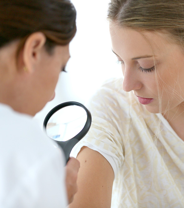
People who work or play in the sun should wear protective clothing, such as a hat and long sleeves. Also, lotion, cream, or gel that contains sunscreen can help protect the skin. Many doctors believe sunscreens may help prevent melanoma, especially those that reflect, absorb, and/or scatter both types of ultraviolet radiation. Sunscreens are rated in strength according to a sun protection factor (SPF). The higher the SPF, the more sunburn protection is provided. Sunscreens with an SPF value of 2 to 11 provide minimal protection against sunburns. Sunscreens with an SPF of 12 to 29 provide moderate protection. Those with an SPF of 30 or higher provide high protection against sunburn. It is advisable to wear a sunscreen that protects against both UVA and UVB. Sunglasses that have UV-absorbing lenses should also be worn. The label should specify that the lenses block at least 99 percent of UVA and UVB radiation.
Early Detection of Melanoma
Because melanoma usually begins on the surface of the skin, it often can be detected at an early stage with a total skin examination by a trained health care worker. Checking the skin regularly for any signs of the disease increases the chance of finding melanoma early. A monthly skin self-exam is very important for people who have any of the known risk factors, but doing skin self-exams routinely is a good idea for everyone. Self-screening was shown in studies to reduce the frequency of advanced lesions in patients with melanoma.
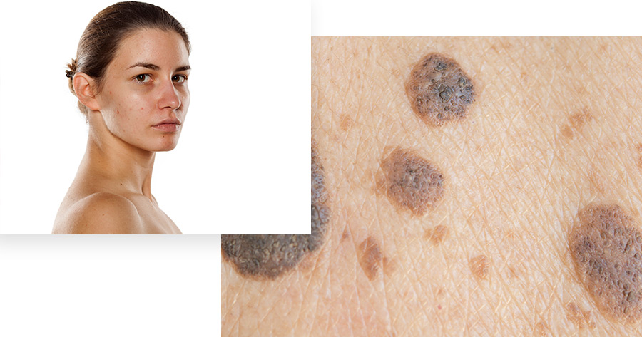
Here is how to do a skin self-exam:
- After a bath or shower, stand in front of a full-length mirror in a well-lighted room. Use a hand-held mirror to look at hard-to-see areas.
- Begin with the face and scalp and work downward, checking the head, neck, shoulders, back, chest, and so on. Be sure to check the front, back, and sides of the arms and legs. Also, check the groin, the palms, the fingernails, the soles of the feet, the toenails, and the area between the toes.
- Be sure to check the hard-to-see areas of the body, such as the scalp and neck. A friend or relative may be able to help inspect these areas. Use a comb or a blow dryer to help move hair so you can see the scalp and neck better.
- Be aware of where your moles are and how they look. By checking your skin regularly, you will become familiar with what your moles look like. Look for any signs of change, particularly a new black mole or a change in outline, shape, size, color (especially a new black area), or feel of an existing mole. Also, note any new, unusual, or “ugly-looking” moles. If your doctor has taken photos of your skin, compare these pictures with the way your skin looks on self-examination.
- Check moles carefully during times of hormone changes, such as adolescence, pregnancy, and menopause. As hormone levels change, moles may change.
- It may be helpful to record the dates of your skin exams and to write notes about the way your skin looks. If you find anything unusual, see your doctor right away. Remember, the earlier a melanoma is found, the better the chance for a cure.
In addition to doing routine skin self-exams, people should have their skin checked regularly by a doctor or nurse specialist. A doctor can do a skin exam during visits for regular checkups. People who think they have dysplastic nevi should point them out without delay to the doctor. It is also important to tell the doctor about any new, changing, or “ugly-looking” moles. It may be preferable in certain high-risk settings such as a history of sunburn, presence of other cutaneous malignancies or melanoma in the past, positive family history of melanoma to see a specialized dermatologist for regular inspections of your skin. Early detection of melanoma may not be easy in individuals having pigmented skin. Although the presence of a degree of skin pigmentation is protective against melanoma, the risk is not zero. The melanoma in such cases develops especially on the hands, feet or nails, but can arise on any other part of the body and on the mucosal surfaces. The patients are encouraged to perform monthly skin self-checks, and to be aware of the criteria used to assess moles. Consult with a dermatologist specializing in pigmented skin lesions and in the detection technique called “dermoscopy” is advisable. Caution for patients with pigmented skin is recommended, because melanoma although less frequent, may be more aggressive and present at an advanced stage.


Distinguishing Benign Moles from Melanoma
To prevent melanoma, it is important to examine your skin on a regular basis, and become familiar with moles, and other skin conditions, in order to better identify subtle changes. Any location on the skin can be involved by melanoma, but the sun exposed areas, and in particular the back and legs are frequently affected. Certain areas can be easily missed on inspection, such as the scalp or the back of the ears.
According to recent research, certain moles are at a higher risk for changing into malignant melanoma. Moles that are present at birth, and atypical moles, have a greater chance of becoming malignant. Frequently, melanomas arise as single lesions. Occasionally, one spot may stand out as looking most atypical, and such lesions would be of particular concern.
Recognizing changes in your moles, by following this ABCD Chart, is crucial in detecting malignant melanoma at its earliest stage. The warning signs are (Table):
Melanomas vary greatly in appearance. Some melanomas may show all of the ABCD characteristics, while other may only show changes in one or two characteristics. Always consult your physician for a diagnosis.
While the ABCD system was designed to allow detection of melanoma by healthcare professionals, other tools have been developed recently.
A simple checklist consisting of seven items was created for the general public. Even one of the following items needs to alert an individual performing self-assessments to consult with a medical professional:
- Changes in shape
- Changes in size
- Changes in color
- Inflammation
- Any lesion greater than 7 millimeters
- Presence of bleeding or crusting
- Presence of inflammation
- Changes in sensation in the area of the lesion
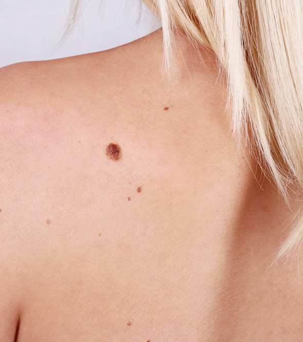

When used correctly, the sensitivity of these criteria is very high, their role being to indicate that a more thorough evaluation and intervention are needed. Since benign non-cancerous lesions may also contain similar features, it is strongly advisable to promptly consult a medical professional in regard to any suspicious lesion. Although a change in size of a mole is the most frequent change associated with melanoma, all the other signs should be given importance. Although not mentioned in these criteria, occurrence of a bump or “vertical growth phase” over a nevus or a new lesion, and presence of pain associated with a pigmented lesion should also be regarded as red flags for melanoma. Bleeding is often associated with thick lesions, therefore the diagnosis is preferable to be made before the occurrence of this symptom in order to ensure early detection.
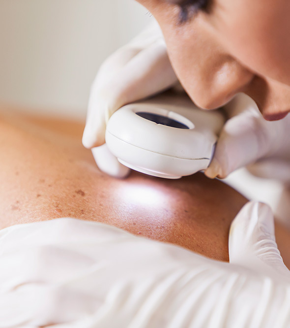
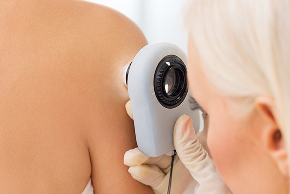
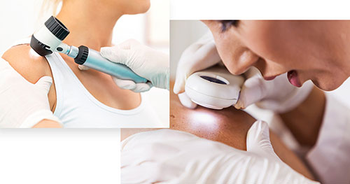
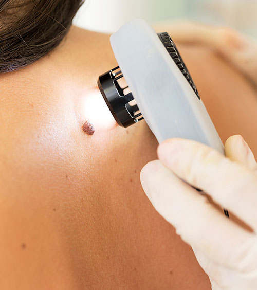
The sensitivity of the seven-item tool appears to be even higher than for the ABCDE criteria. Recognition of melanoma is considered to be highest when the exam is conducted by trained dermatologists, but it is not reaching 100%. In order to increase the accuracy of clinical examination and to help distinguish melanoma from other skin entities, a new advance employes the use of dermoscopy. This technique uses an optical magnifying instrument which is applied on the skin and allows better visualization of the structural details through a layer of alcohol or oil immersion. A light source and magnification in the range of 10x allow visualization of details which are not reachable by naked eye inspection. Dermoscopy is becoming increasingly used among dermatologists and results in an increased number of lesions diagnosed early, along with a decrease in the number of surgical biopsies which would be performed because of uncertainties in diagnosis. The sensitivity and the ability to correctly identify melanomas was shown to be improved compared to naked eye examination. Dermoscopy has been successfully applied in patients with dark skin, leading to early and accurate diagnosis of melanoma.
Total body mole maping consists of pictures taken with adequate resolution so that pictures taken at one visit may be compared with the appearance of the mole at the next visit. Its value applies to patients with more than 200 nevi, and instances of high-risk large and otherwise suspicious lesions. Comparison with the baseline photographs allows detection of new moles, assessment of enlarging lesions, and evaluation of changes in the aspect of pre-existing nevi.
Based on the physical changes and on the history taken from the patient, a doctor decides that a mole should be removed so that the tissue can be examined under a microscope. A doctor may want to watch a slightly abnormal mole closely to see whether it changes over time. The removal of a mole, called a biopsy, is usually done in the doctor’s office using a local anesthetic. It generally takes only a few minutes. The patient may require stitches, and a small scar will remain after healing. A pathologist examines the tissue under a microscope to see whether the melanocytes are normal, dysplastic, or cancerous.


Seeking Medical Advice
Sometimes it is necessary to see a specialist. A dermatologist (skin doctor) have the most training in diseases of the skin. Our doctors at Advanced Dermatology PC are participants of the AAD annual melanoma screening sessions and are often cited in media for their expertise in skin cancer prevention and treatment.
Melanoma may run in families, and members of these families are at high risk for the disease. In some of these families, certain members also have a large number (usually over 100) of dysplastic nevi. These people have an especially high risk of developing melanoma. When two or more family members develop melanoma, it is important for all of the patients’ close relatives (parents, brothers, sisters, and children above the age of 10) to see a doctor and be examined carefully for dysplastic nevi or any signs of melanoma. The doctor can then decide how often each person needs to be seen. (Doctors may recommend that these family members have checkups every 6 months.) Anyone who has a large number of dysplastic nevi also should be examined regularly.
Because most moles, including most dysplastic nevi, do not develop into melanoma, removing all of them is not necessary. In fact, a phenomenon of benign involution if frequently observed in nevi. A doctor can recommend when and when not to remove moles. Usually, only moles that look like melanoma, those that change, or those that are both new and look abnormal need to be removed. Prophylactic removal of benign nevi is therefore not routinely recommended, however this is sometimes considered for cosmetic and psychological reasons.
Genetic Features of Melanoma
One tenth of melanomas are considered to be familial, meaning that they affect several family members. If a first-degree relative is affected by melanoma, the risk increases by 10 times. If in addition to having a first-degree relative with melanoma the patient also has atypical (“dysplastic”) nevi, the risk is much increased. The presence of multiple cutaneous melanomas and multiple nevi in one individual can also be associated with the presence of familial melanoma. While up to one quarter of familial cases are associated with genetic mutations in a gene called CDKN2A, only approximately 1% of isolated (nobody else in the family having melanoma) melanomas contain this abnormality. Genetic testing may be considered for families where at least two members have melanoma, melanoma occurring at young ages, or presence of pancreatic cancer in other family members. The decision regarding genetic testing must be taking after discussing with your physician and having a genetic counseling session, which will inform you regarding the potential implications of the information generated by such testing.
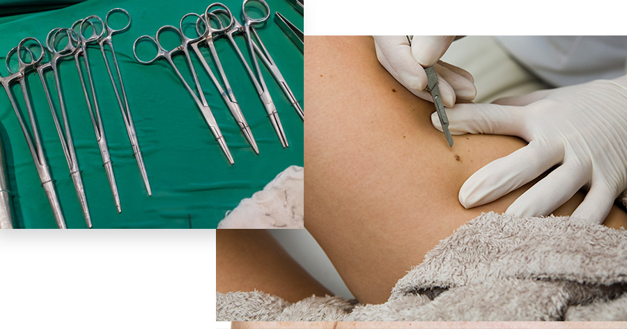

Suspicious lesions are removed surgically. The removal of the entire mole or a sample of tissue for examination under a microscope is called a biopsy. It is considered preferable to remove the lesion entirely (“excisional biopsy”) with no cancer cells present at the margins of the piece extracted. However, for large lesions or melanomas in difficult anatomic locations a smaller part is removed (“incisional biopsy”). Although not removing the whole lesion from the beginning is not considered to impact the outcome as long as a full resection is done subsequently, very superficial biopsies are not generally recommended because they can potentially miss a deep melanoma.
Analysis of microscopic features of the excised specimens is performed routinely to confirm the diagnosis of melanoma and distinguish from other pigmented lesions that may appear very similar. Although many of these non-melanoma entities are benign, some of them are considered “atypical” or “dysplastic” variants, which place them at increased risk and mandate surgical excision and surveillance.
Adjuvant Treatment
“Adjuvant treatment” represents further therapy addressed to reduce the risk of recurrence and possibly improving the survival of some patients in certain high-risk groups. Although a precise determination of the best strategy can only be made by a specialized physician, patients having lymph node involvement by melanoma and those having advanced skin lesions should expect to have a discussion regarding such treatments. Several modalities are available currently, with interferon-alpha and vaccines being the most commonly employed. An alternative strategy, used especially for lower risk lesions is to provide clinical surveillance with no active treatment. Your physician may recommend one of these treatments, or an investigational approach depending on the assessed risk of the skin lesion, the age and physical parameters of the patient, and availability of certain protocols. Although being the most extensively studied and used, interferon-alpha is not universally recognized as a standard of care for adjuvant treatment for melanoma.
Treatment for Metastatic Disease
Treatment for “metastatic disease” or disease that has spread at distance is complex and involves modalities such as biological agents (interleukin-2, interferon-alpha, antibodies against CTLA4, vaccines, etc.), chemotherapy, radiation therapy, surgery, or integrated approaches.


Stages of Melanoma
After reaching a diagnosis of melanoma, staging or assessment of the extend of the disease is performed in an effort to predict the outcome and the need for other studies and further treatment.
Staging means determining the extent of the disease. More evolved cutaneous lesions have an increased chance to spread to the lymph nodes or to distant sites inside the body. Any organ can become involved with melanoma, but more frequently involved are skin and subcutaneous tissues, lungs, liver, and brain.
Features that determine the staging include the skin tumor thickness, presence of ulceration and microscopic islands surrounding the main tumor, and the presence and number of lymph nodes containing melanoma cells. Only a fraction of patients have melanoma involving regional lymph nodes or distant metastases at diagnosis, which place them in an advanced stage (stages III and IV, respectively).
Stage Ia
Consists of a very thin tumor (< 1 mm) without ulceration.
Stage Ib
Consists of the same thickness of a tumor but having ulceration or invading to a Clark level III or IV, or a thin tumor (1.01 – 2 mm) without ulceration.
Stage IIa
Comprises thin tumors (1.01 – 2 mm) with ulceration and intermediate thickness tumors (2.01 – 4 mm) without ulceration.
Stage IIb
Comprises intermediate thickness tumors (2.01 – 4 mm) with ulceration, or thick melanomas (> 4 mm) without ulceration.
Stage IIc
Consists of thick lesions (> 4 mm) with ulceration.


Stage III
Melanoma is diagnosed when the tumor was found inside the lymph nodes that drain the territory of the melanoma lesion, either as micrometastases (can only be found by examination under the microscope) or macrometastases (can be appreciated by manual palpation during a clinical exam), or other deposits of melanoma are found along the trajectory of lymphatic channels (“in-transit metastases” or “satellites”).
Stage IV
Melanoma consists of distant spread to sites such as lymph nodes, skin, or under the skin (Stage M1a), lungs (Stage M1b), or any other distant organ (Stage M1c).
The prognosis, or chances of melanoma to progress to advanced stages and become life-threatening are influenced by certain factors, such as:
- a larger thickness of the lesion (measured by the so-called Breslow dept in millimeters), as shown above, is associated with a higher stage, which makes the prognosis less favorable.
- Clark’s invasion level (on a scale form I to IV) is the anatomic layer of the skin to which the tumor reaches. The higher the number, the deeper the lesion invades, which signifies a less favorable outcome. Clark level I means that the melanoma is only located in the epidermis, the most superficial layer of the skin. Clark level II signifies that the melanoma invaded the next layer called papillary dermis. Clark level III is reached when the tumor reaches but does not invade the reticular dermis. Clark level IV means that the tumor reached into the reticular or the deep dermis. Clark level V is reached when the subcutaneous layer is involved with melanoma.
- Lymph nodes or distant organs being involved with melanoma.
- The advanced age and male sex have been associated with a less favorable outcome


Once Melanoma Has Been Diagnosed
Assessment of whether the lymph nodes are involved with melanoma is made depending on certain characteristic of the melanoma lesion on the skin. If the nodes are large enough to be found during a physical exam performed by your physician, then a referral will be generally made to a surgeon for a complete surgical extraction of lymph nodes in that territory. If lymph nodes are not detested clinically, but the melanoma has certain high-risk features as assessed by your dermatologist or by a medical oncologist, a specialized surgeon will perform a procedure called “sentinel lymph node biopsy”. This procedure consists of injection of blue dye and a radioactive tracer around the site of the primary skin lesion, after it has been removed. The lymph node uptaking these tracing substances will be located and removed surgically. The advantage of this procedure which now is used routinely is that it produces less risk of postoperative complications (in particular “lymphedema” or chronic swelling of an extremity), while producing similar results as a complete lymphatic removal. If the “sentinel lymph node” is found to be involved with melanoma by looking under the microscope and performing other very sensitive tests, than all the lymph nodes in the region that drain the primary melanoma need to be removed. The best course of action is usually determined for an individual patient by a multidisciplinary team composed of a dermatologist, a medical oncologist, and a surgeon.
Depending on the assessed risk for distant spreading of an individual skin lesion, the physician may use in some patients chest X-rays, CT scans, MRIs, and/or PET scans in addition to a detailed physical exam to evaluate the rest of the body. These studies may be repeated at regular intervals, but their frequency typically decreases as time passes after the diagnosis of the skin melanoma. While being on such a surveillance program, the patient needs to report promptly to the physician any signs or signs of dysfunction including, but not limited to shortness of breath, cough, weakness in any part of the body, seizures, abnormalities in perception or thinking, or pains in any location.
A full excision of the tumor is performed, with removal of a portion of normal skin where isolated melanoma cells may possibly reside. These “wide margins” of removal depend on the thickness of the primary melanoma lesion:
– for melanoma which are very superficial (labeled “in-situ”), a 5 milimiter margin around the tumor was determined appropriate.
-for melanomas 0-1 cm, a 1 cm margin is used.
– for melanomas of 1-2 mm, a 1-2 cm margin is recommended.
-for melanomas greater than 2 mm, a 2 cm margin is utilized.
Of note, significantly larger margins were used in the past, but they were not shown to improve the outcome, while leaving larger scars. Determining the optimal margin may involve other considerations such as the anatomical location, and cosmetical needs of an individual patient. Excision of melanomas located on the lips, nose, ears, eyelids, hands or feet may require a referral to a plastic surgeon for best cosmetic and functional outcomes after the resection. A thorough medical evaluation is followed by a discussion between the medical team and the pacient in regard to the method utilized.


Knowledge of the above information and a few simple changes in behavior and lifestyle can prevent skin cancer:
- Wear sun-protective clothing during peak sun hours (10a.m – 2p.m).
- Use sunscreen with both UVA/UVB & physical blocks. Several studies have demonstrated that the use of sun-protective wear and sunscreen can decrease the number of moles and premalignant lesions.
- The American Academy of Dermatology recommends broad-spectrum sunscreen covering both UVA & UVB with a Sun Protection Factor (SPF) of at least 15 or above with reapplication every 2 hours when outdoors, even on cloudy days.
- Make efforts to minimize sun exposure and to apply sunscreen to children aged 6 months and older.
- Clinical trials have demonstrated that sunscreens are effective in reducing the incidence of actinic keratoses and squamous cell carcinomas.
- Another randomized trial demonstrated that, among children who are at high risk for developing melanoma, sunscreens are effective in reducing moles, the precursors and strongest risk factor for melanoma.
- It is strongly recommended to avoid tanning salons due to its possible association with skin cancers.
